&geometry(249x120) "Recover Radiology")
Posted in Ultrasound
What Does a Sonographer Do?
 "What Does a Sonographer Do?")
When you arrive for an ultrasound appointment, the friendly professional who scans your abdomen, shoulder, or heart isn't a doctor—they're a sonographer, a highly trained specialist in ultrasound imaging. Many patients are unsure about the sonographer's role and how they fit into the diagnostic imaging team. Understanding who sonographers are, what they do, and how they contribute to your diagnostic journey can help you feel more confident during your ultrasound appointment.
Who Is a Sonographer?
A sonographer is a healthcare professional who specializes in using ultrasound equipment to produce images of internal structures within the body. They are the individuals who perform ultrasound scans, positioning the ultrasound probe over the area being examined, adjusting settings, and capturing the images that allow radiologists to make diagnoses. Sonographers are different from doctors and radiologists—they're imaging specialists focused specifically on ultrasound technology and technique. They work under the direction of radiologists who interpret the images and provide the clinical diagnosis.
Sonographer Qualifications and Training
Becoming a sonographer requires formal education. Most sonographers complete either a Bachelor's degree in Medical Sonography or a Graduate Diploma in Medical Sonography, typically taking 2-3 years depending on the program structure. This education covers human anatomy, physiology, ultrasound physics, scanning techniques, image interpretation, patient communication, and clinical safety. Beyond formal education, sonographers must achieve accreditation from the Australasian Society for Ultrasound in Medicine (ASUM) or the Australian Sonographers Association (ASRA), which involves passing examinations that verify their knowledge and practical competency. Many sonographers pursue additional qualifications and credentials throughout their careers to deepen their expertise in specific ultrasound areas.
What Sonographers Do Day-to-Day
The sonographer's role involves much more than simply moving an ultrasound probe across your skin. They begin by reviewing your clinical referral to understand why imaging is needed and what specific questions the radiologist needs answered. They introduce themselves, explain what to expect during the scan, answer your questions, and help you feel comfortable. During the scan, they use specialized ultrasound equipment to acquire images of internal structures, constantly adjusting probe position, angle, depth, and settings to capture the clearest possible images. They recognize normal and abnormal findings, identify which images are most diagnostically useful, and acquire multiple views to give the radiologist complete information. After scanning, they ensure all images are properly labeled, stored securely, and passed to the radiologist for interpretation
Sonographer Specialisations
While some sonographers are general sonographers skilled across multiple body areas, many specialize in specific ultrasound fields. General sonographers perform abdominal, pelvic, and other routine ultrasounds. Cardiac sonographers specialize in heart ultrasound, examining heart function and structure in detail. Vascular sonographers focus on blood vessels, assessing blood flow and looking for clots or other abnormalities. Obstetric sonographers work with pregnant patients, monitoring fetal development and assessing pregnancy health. Musculoskeletal sonographers specialize in imaging joints, tendons, ligaments, and muscles—useful for sports injuries and joint pain. Each specialisation requires additional training and expertise within that particular area, allowing sonographers to develop deep knowledge of their specialty.
Different Ultrasound Modalities
Modern ultrasound technology allows sonographers to perform increasingly sophisticated scanning. Standard ultrasound produces real-time, cross-sectional images. Doppler ultrasound assesses blood flow and direction, helping identify clots or abnormal flow patterns. 3D and 4D ultrasound creates three-dimensional images, particularly valuable in obstetrics. Elastography measures tissue stiffness, useful for assessing liver fibrosis or muscle injury. Contrast-enhanced ultrasound uses microbubbles to enhance tissue visualization. Sonographers must understand when each modality is appropriate, how to use the technology effectively, and how to interpret the resulting images.
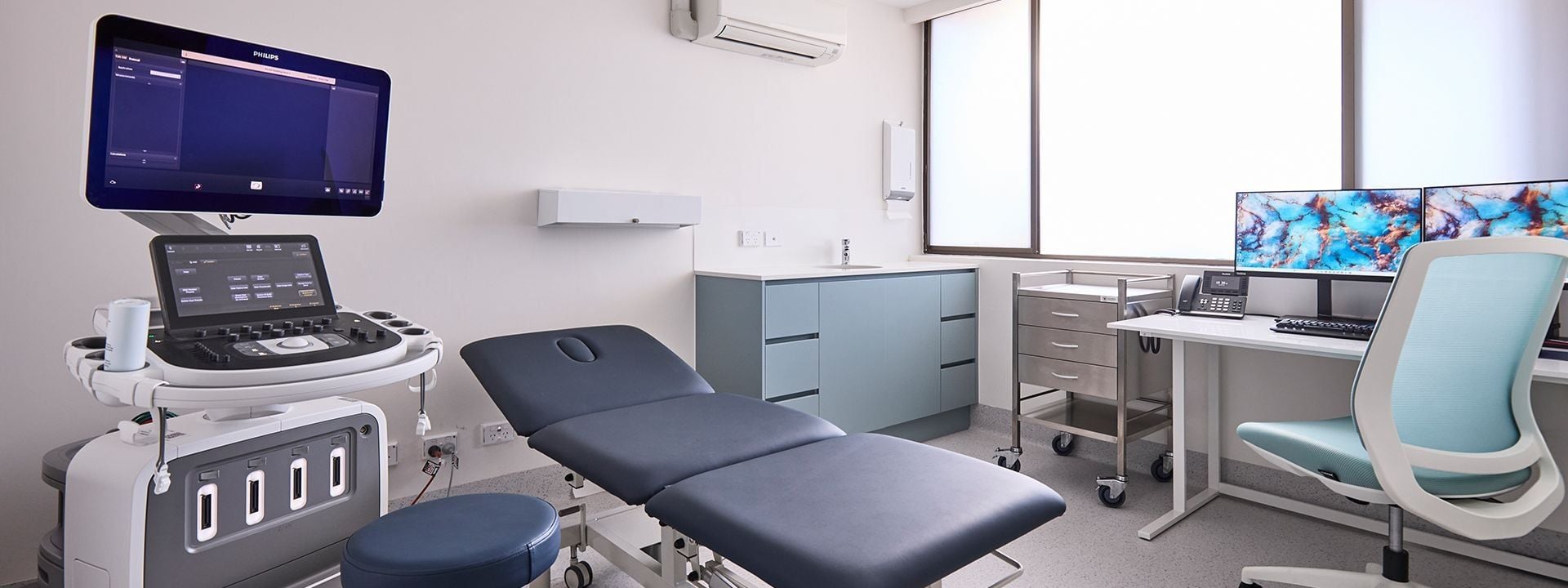
Recover Radiology Sonographers
At Recover Radiology in Morphett Vale, our sonographers bring extensive experience and specialized expertise to every ultrasound appointment. With over 25 years of combined experience, our team has developed the skills to perform high-quality ultrasound imaging across a range of clinical indications. They're trained professionals who take pride in patient care, image quality, and contributing to accurate diagnoses that guide your healthcare. When you attend an ultrasound appointment with us, you're working with experienced specialists committed to providing you with excellent diagnostic imaging.
Book Your Ultrasound Today
If your GP has recommended an ultrasound, our skilled sonographers at Recover Radiology are ready to provide you with excellent imaging care. Call us at 08 7081 3078 to book your appointment. We're located at 1-3 Doctors Road, Morphett Vale, SA 5162, open Monday to Friday, 8:30am to 5:30pm. Ultrasound imaging is safe, radiation-free, and provides real-time diagnostic information. Our experienced sonographers and radiologists work together to ensure accurate diagnoses that guide your healthcare.
| Posted in:Recover RadiologyBulk BillingUltrasound |